
NEWS!
- 25.09.2018: "Graph Saliency Maps Through Spectral Convolutional Networks: Application to Sex Classification with Brain Connectivity" by Salim Arslan et al. was awarded the GRAIL Best Poster Award
- 25.09.2018: "Multi-modal Disease Classification in Incomplete Datasets Using Geometric Matrix Completion" by Gerome Vivar et al. was awarded the GRAIL Best Paper Award
- 25.09.2018: GRAIL LNCS Proceedings are already available at: https://link.springer.com/book/10.1007/978-3-030-00689-1
- 18.09.2018: The program has been updated given to the cancellation of 3 talks due to Visa issues.
- 20.07.2018: The program of the workshop is now available:
- 20.07.2018: We extended the deadline for abstract submissions to 27th July!
- 12.06.2018: Our 2nd Keynote Speaker will be Prof. Dimitri Van de Ville talking about graph signal processing for human brain imaging.
- 06.06.2018: Deadline for complete papers is extended until 18th June!
- 21.05.2018: CMT System is open for submissions of both, complete and abstract papers! Click here to submit your manuscript
- 21.05.2018: PC Members are online! (More TBA)
- 09.04.2018: This year we will accept papers in two modalities: Complete papers (8-10 pages) and Abstract papers (2-3 pages). Read more about it in the section "Submit a paper"
- 09.04.2018: GRAIL accepted papers will be invited to submit an extended version to a Special Issue on Graphs in Biomedical Image Analysis that we will organize in a top-tier journal of the area.
- 09.04.2018: The GRAIL Best Paper Award will include a cash price sponsored by EntelAI and MedIAN!
- 30.03.2018: The GRAIL Website is up and running!


SCOPE

GRAIL 2018 is the second international workshop on GRaphs in biomedicAl Image anaLysis, organised as a satellite event of MICCAI 20178 in Granada, Spain.
Graph-based models have been developed for a wide variety of problems in computer vision and biomedical image analysis. Applications ranging from segmentation, registration, classification, and shape modelling, to population analysis have been successfully encoded through graph structures, demonstrating the versatile and principled nature of the graph based approaches.
Graphs are powerful mathematical structures which provide a flexible and scalable framework to model objects and their interactions in a readily interpretable fashion. As a consequence, an important body of work has been developed around different methodological aspects of graph including, but not limited to, graphical models, graph-theoretical algorithms, spectral graph analysis, graph dimensionality reduction, and graph-based network analysis. However, new topics are also emerging as the outcome of interdisciplinary studies, shedding light on areas like deep structured models and signal processing on graphs.
With this workshop we aim to highlight the potential of using graph-based models for biomedical image analysis. Our goal is to bring together scientists that use and develop graph-based models for the analysis of biomedical images and encourage the exploration of graph-based models for difficult clinical problems within a variety of biomedical imaging contexts.
The covered topics include but are not limited to:
- Probabilistic graphical models for biomedical image analysis
- Discrete and continuous optimization for graphical models
- Signal processing on graphs for biomedical image analysis
- Deep/machine learning on structured and unstructured graphs
- Convolutional neural networks on graphs
- Graphs for large scale population analysis
- Graph-based shape modeling and dimensionality reduction
- Combining imaging and non-imaging data through graph structures
- Graph-based generative models for biomedical image analysis
- Graph spectral methods
- Algorithms on graphs
- Graphs in neuroimaging
- Applications of graph-based models and algorithms to biomedical image analysis tasks (segmentation, registration, classification, etc.)
Program
| 08:30 – 09:30 | Registration |
| 09:30 – 09:45 | Welcome and opening remarks |
| 09:45 – 10:30 | Keynote Speaker 1: Michael Bronstein "Deep learning on Graphs" |
| 10:30 – 11:00 | Session 1: Graph CNNs
|
| 11:00 – 11:30 | Coffee Break |
| 11:30 – 12:15 | Keynote 2: Dimitri Van de Ville "Graph Signal Processing Opens New Perspectives for Human Brain Imaging" |
| 12:15 – 13:30 | Session 2: Graph based models for biomedical applications
|
| 13:15 – 13:30 | Best paper award and closing remarks |
| 13:30 – 15:00 | Poster session and lunch |
Keynote Speakers
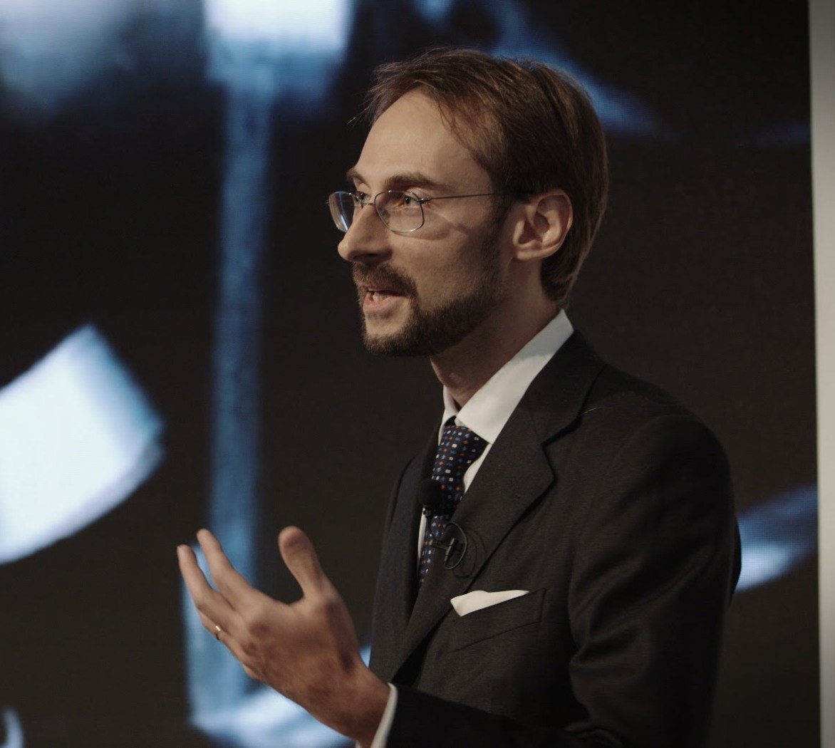
Prof. Michael Bronstein
Imperial College / USI Lugano
Deep Learning on Graphs
Bio : Michael Bronstein (PhD with distinction 2007, Technion, Israel) is a professor at USI Lugano, Switzerland and Imperial College London, UK where he holds the Chair in Machine Learning and Pattern Recognition and Royal Society Wolfson Merit Award. During 2017-2018 he was a fellow at the Radcliffe Institute for Advanced Study at Harvard University. Michael's main research interest is in theoretical and computational methods for geometric data analysis. He authored over 150 papers, the book Numerical geometry of non-rigid shapes (Springer 2008), and over 20 granted patents. He was awarded four ERC grants, two Google Faculty Research awards, Amazon AWS Machine Learning award, and Rudolf Diesel fellowship (2017) at TU Munich. He was invited as a Young Scientist to the World Economic Forum, an honor bestowed on forty world’s leading scientists under the age of forty. Michael is a Senior Member of the IEEE, alumnus of the Technion Excellence Program and the Academy of Achievement, ACM Distinguished Speaker, and a member of the Young Academy of Europe. In addition to academic work, Michael is actively involved in commercial technology development and consulting to start-up companies. He was a co-founder and technology executive at Novafora (2005-2009) developing large-scale video analysis methods, and one of the chief technologists at Invision (2009-2012) developing low-cost 3D sensors. Following the multi-million acquisition of Invision by Intel in 2012, Michael has been one of the key developers of the Intel RealSense technology in the role of Principal Engineer.
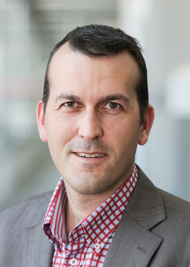
Prof. Dimitri Van De Ville
EPFL / University of Geneva
Graph Signal Processing Opens New Perspectives for Human Brain Imaging
Abstract: State-of-the-art magnetic resonance imaging (MRI) provides unprecedented opportunities to study brain structure (anatomy) and function (physiology). Based on such data, graph representations can be built where nodes are associated to brain regions and edge weights to strengths of structural or functional connections. In particular, structural graphs capture major neural pathways in white matter, while functional graphs map out statistical interdependencies between pairs of regional activity traces. Network analysis of these graphs has revealed emergent system-level properties of brain structure or function, such as efficiency of communication and modular organization. In this talk, graph signal processing (GSP) will be presented as a novel framework to integrate brain structure, contained in the structural graph, with brain function, characterized by activity traces that can be considered as time-dependent graph signals. Such a perspective allows to define novel meaningful graph-filtering operations of brain activity that take into account the anatomical backbone. For instance, we will show how activity can be analyzed in terms of being aligned versus liberal with respect to brain structure, or how additional prior information about cognitive systems can be incorporated. The well-known Fourier phase randomization method to generate surrogate data can also be adapted to this new setting. Finally, recent work will highlight how the spatial resolution of this type of analyses can be increased to the voxel level, representing a few ten thousands of nodes.
Bio: Dimitri Van De Ville (Senior Member, IEEE) received the M.S. degree in engineering and computer sciences and the Ph.D. degree from Ghent University, Ghent, Belgium, in 1998, and 2002, respectively. After a postdoctoral stay (2002-2005) at the lab of Prof. M. Unser at the Ecole Polytechnique Fédérale de Lausanne (EPFL), Lausanne, Switzerland, he became responsible for the Signal Processing Unit at the University Hospital of Geneva, Geneva, Switzerland, as part of the Centre d'Imagerie Biomédicale (CIBM). In 2009, he received a Swiss National Science Foundation professorship and since 2015 became Professor of Bioengineering at the EPFL and the University of Geneva, Geneva, Switzerland. His research interests include wavelets, sparsity, graphs, pattern recognition, and their applications in computational neuroimaging.
Dr. Van De Ville served as an Associate Editor for the IEEE Transactions on Image Processing from 2006 to 2009 and the IEEE Signal Processing Letters from 2004 to 2006, as well as Guest Editor for several special issues. He was the Chair of the Bio Imaging and Signal Processing (BISP) Technical Committee of the IEEE Signal Processing Society (2012-2013) and is the Founding Chair of the EURASIP Biomedical Image & Signal Analytics SAT. He is Co-Chair of the biennial Wavelets & Sparsity series conferences, together with V. Goyal, Y. Lu, and M. Papadakis. He was a recipient of the Pfizer Research Award 2012, the NARSAD Independent Investigator Award 2014, and the Leenaards Foundation Award 2016.
Submit a paper
Submissions should be done through the GRAIL CMT portal. The submission system will open on May 20th.
This year we will accept papers in 2 formats:
- Complete papers: Papers describing original research with length of 8-10 pages. These papers are eligible for the "Best Paper Award" and will be published in the GRAIL Open Proceedings and Lecture Notes in Computer Sciences (LNCS) series. Submissions should be anonymous, and formatted following the LNCS Style. All complete papers will be peer-reviewed by 3 members of the program committee, with double-blinded review process. The selection of the papers will be based on significance of results, technical merit, relevance and clarity of presentation.
- Abstracts: In order to encourage interesting discussions within the community, this year we are also inviting papers describing on-going, recently published or original research in a short paper of 2-3 pages. Depending on the number of submissions, we will organize a poster or oral session during GRAIL, where accepted short papers will be invited to present. The selection of the short abstracts will be made by members of the program committee and/or organizers. Submissions should be anonymous, and formatted following the LNCS Style.
GRAIL Best Paper Award and Special Issue
The authors of the best paper of the workshop will receive a cash price (sponsored by EntelAI and MedIAN).
Accepted papers will be invited to submit an extended version to a Special Issue on Graphs in Biomedical Image Analysis in a top-tier journal of the area, that we will organize after the event.
Important dates
| 20 May 2018 | Submission system opens. |
| Complete Paper submission deadline. | |
| 10 July 2018 | Reviews due. |
| Notification of acceptance. | |
| 20 July 2018 | Camera-ready papers (Complete papers). |
| Abstract Paper submission deadline. | |
| 3rd August 2018 | Notification of acceptance for Abstract Paper submission. |
Organising Committee
- Enzo Ferrante, CONICET / Universidad Nacional del Litoral, eferrante (at) sinc.unl.edu.ar
- Sarah Parisot, AimBrain, Sarah (at) aimbrain.com
- Aristeidis Sotiras, University of Pennsylvania, aristeidis.sotiras (at) uphs.upenn.edu
- Bartlomiej Papiez, University of Oxford, bartlomiej.papiez (at) bdi.ox.ac.uk
Program Committee
- Kayhan Batmanghelich, University of Pittsburgh / Carnegie Mellon University, US
- Eugene Belilovsky, INRIA / KU Leuven, France
- Siddhartha Chandra, CentraleSupelec / INRIA, France
- Xin Chen, The University of Nottingham, UK
- Emilie Chouzenoux, INRIA Saclay, France
- Puneet K. Dokania, Oxford University, UK
- Ben Glocker, Imperial College London, UK
- Ali Gooya, University of Sheffield, UK
- Mattias Heinrich, University of Luebeck, Germany
- Lisa Koch, ETH Zurich, Switzerland
- Evgenios Kornaropoulos, University of Cambridge, UK
- Sofia Ira Ktena, Imperial College London, UK
- Georg Langs, University of Vienna / MIT, Austria, Austria
- Jose Ignacio Orlando, Medical University of Vienna, Austria
- Yusuf Osmanlioglu, University of Pennsylvania, US
- Yangming Ou, Harvard University, US
- Nikos Paragios, CentraleSupelec / INRIA, France
- Sotirios Tsaftaris, University of Edinburgh, UK
- Maria Vakalopoulou, Université Paris-Saclay / INRIA, France
- William Wells III, Harvard Medical School, US
Sponsors